Expanded Nursing Uganda Explanation
Cardiovascular system should be understood beyond a short definition. Link the concept to patient history, focused assessment, common risks, nursing priorities, documentation and evaluation of outcomes.
Contents — 43 sections (tap to expand)
01 Topic: Structures and functions of various body systems - Cardiovascular System (PEX 1.8.1)
The Cardiovascular system is the transport system of the body, through which the nutrients are conveyed to places where these are utilized, and the metabolites (waste products) are conveyed to appropriate places from where these are expelled.
The conveying medium is a liquid tissue, the blood, which flows in tubular channels called blood vessels. The circulation is maintained by the central pumping organ called the heart.
02 The Heart
The heart is a small muscular hollow organ—roughly the size of your fist. It is located in mediastinum near the anterior chest wall, directly posterior to the sternum. The mediastinum contains the great vessels, which are attached at the base of the heart, as well as the thymus, esophagus, and trachea.
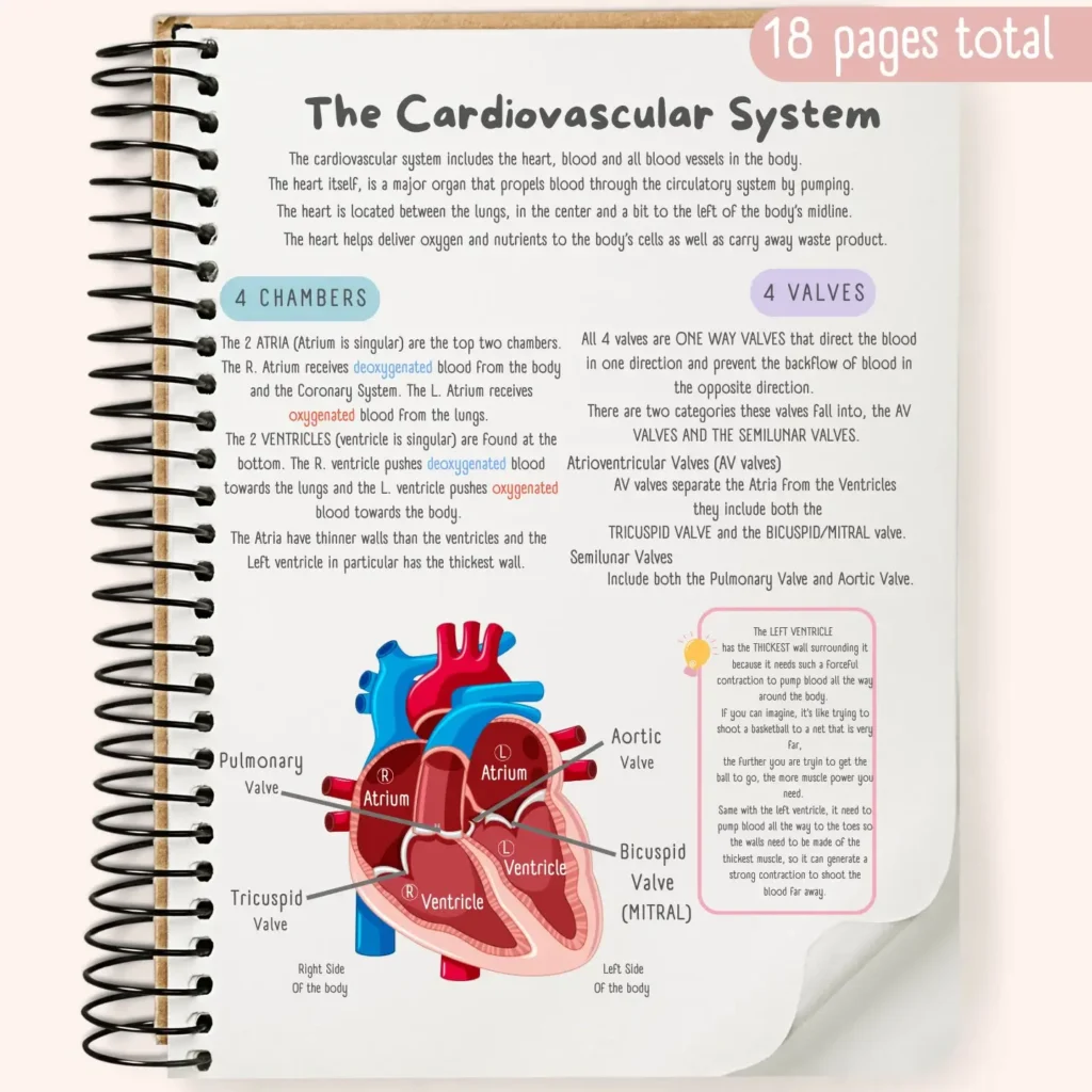
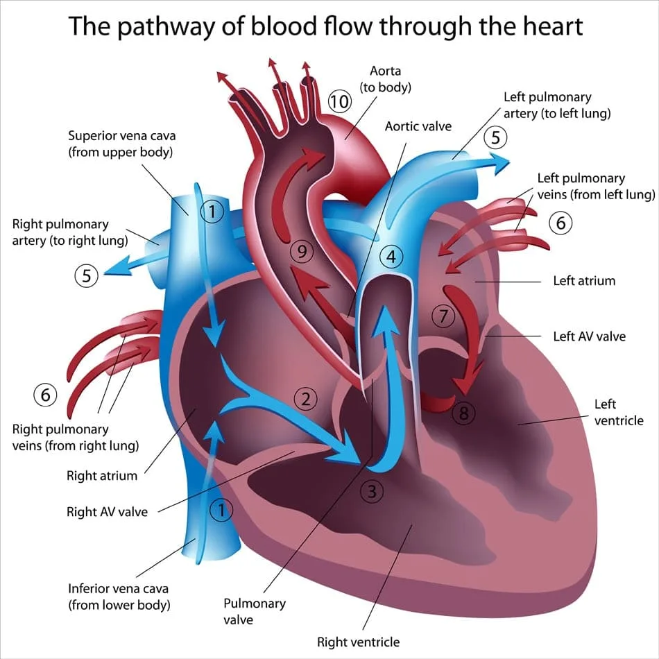
The heart has four muscular chambers, the right and left atria and right and left ventricles . These four chambers work together, pumping blood through a network of blood vessels that connect the heart to peripheral tissues.
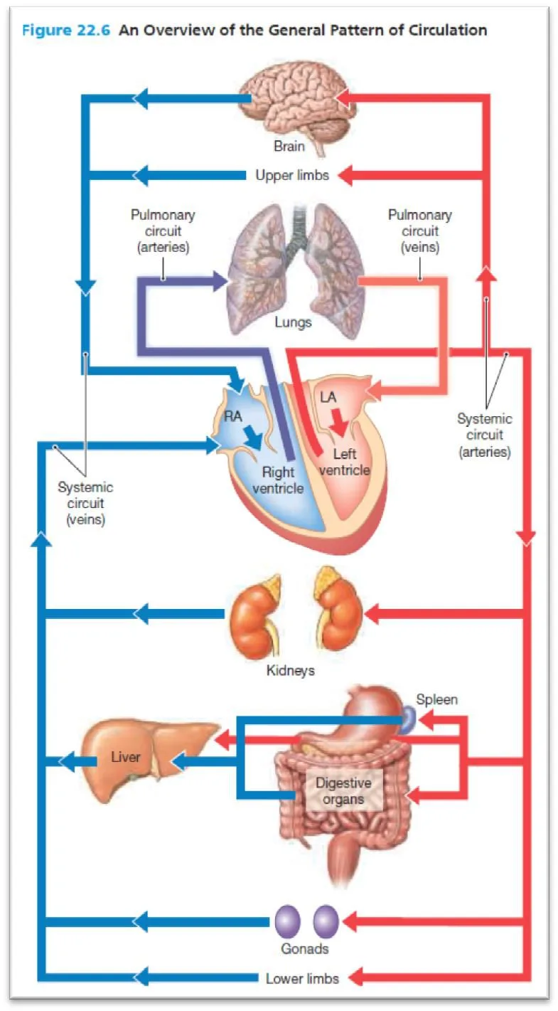
The network of vessels is divided into two circuits: the pulmonary circuit and the systemic circuit . The pulmonary circuit carries carbon dioxide–rich blood from the heart to the gas exchange surfaces of the lungs and returns oxygen- rich blood to the heart. The systemic circuit transports oxygen–rich blood from the heart to the rest of the body’s cells and returns carbon dioxide-rich blood back to the heart. The right atrium receives blood from the systemic circuit, and the right ventricle pumps blood into the pulmonary circuit. The left atrium receives blood from the pulmonary circuit, and the left ventricle pumps blood into the systemic circuit.
With each heartbeat, the atria contract first, followed by the ventricles. The two ventricles contract at the same time and eject equal volumes of blood into the pulmonary and systemic circuits.
Each circuit begins and ends at the heart, and blood flows through these circuits in sequence. Thus, blood returning to the heart from the systemic circuit must complete the pulmonary circuit before re-entering the systemic circuit. The blood vessels of both circuits are arteries, veins, and capillaries.
The Cardiovascular system , also called the circulatory system, is like the body's transport network. It is responsible for moving essential substances throughout the body.
03 Main Parts:
This system has two main parts:
- The heart , which acts as the pump.
- Blood vessels (arteries, veins, capillaries), which are like the tubes or pipes that carry the blood.
04 Function:
The main job of the cardiovascular system is to circulate blood to all parts of the body. Blood carries:
- Oxygen from the lungs to the body tissues.
- Nutrients (like sugar, amino acids) from the digestive system to the tissues.
- Hormones from endocrine glands to their target organs.
- Waste products (like carbon dioxide, urea) from the tissues to organs that remove them (lungs, kidneys, liver).
- Heat to help maintain body temperature.
- Cells and substances that protect the body (part of the immune system).
05 The Heart (The Pump)
The heart is a muscular organ located in the chest, between the lungs, slightly towards the left side. It is about the size of your fist. The heart works continuously, pumping blood throughout your entire life.
06 Structure of the Heart:
The heart is divided into four chambers.
- Two upper chambers called atria (singular: atrium). The right atrium receives blood from the body, and the left atrium receives blood from the lungs.
- Two lower, thicker, muscular chambers called ventricles . The right ventricle pumps blood to the lungs, and the left ventricle pumps blood to the rest of the body.
A wall called the septum separates the right side of the heart from the left side, ensuring blood from the two sides does not mix.
The heart also has valves between the chambers and at the exits of the ventricles. These valves act like one-way doors, making sure blood flows in the correct direction through the heart. When the heart beats, these valves open and close.
07 How the Heart Pumps:
The heart muscle contracts and relaxes in a repeating cycle called the cardiac cycle or heartbeat.
- Systole: This is when the heart muscle contracts and pushes blood out of the chambers.
- Diastole: This is when the heart muscle relaxes and the chambers fill with blood.
The right side of the heart receives blood from the body and pumps it to the lungs. The left side of the heart receives blood from the lungs and pumps it to the body. (This happens at the same time).
08 Blood Vessels (The Pipes)
There are three main types of blood vessels:
09 Arteries:
These vessels carry blood away from the heart .
- They usually carry oxygenated blood (blood rich in oxygen), except for the pulmonary artery, which carries deoxygenated blood to the lungs.
- Arteries have thick, muscular, and elastic walls to withstand the high pressure of blood pumped directly from the heart.
- Large arteries branch into smaller arteries, which then branch into even smaller vessels called arterioles . Arterioles control blood flow into the tiny capillaries.
10 Veins:
These vessels carry blood towards the heart .
- They usually carry deoxygenated blood (blood low in oxygen), except for the pulmonary veins, which carry oxygenated blood from the lungs to the heart.
- Veins have thinner walls and carry blood at lower pressure than arteries.
- Many veins, especially in the arms and legs, have valves inside them. These valves help prevent blood from flowing backwards, especially against gravity. Muscle contractions around the veins also help push blood towards the heart (this is called the skeletal muscle pump ).
- Tiny veins, called venules , collect blood from the capillaries and merge to form larger veins.
11 Capillaries:
These are the smallest blood vessels , connecting arterioles and venules. They form extensive networks within tissues.
Function: This is where the real work of exchange happens! Capillary walls are very thin (only one cell layer thick), allowing oxygen, nutrients, and other substances to pass out of the blood into the surrounding tissues. At the same time, waste products (like carbon dioxide) pass from the tissues into the blood through the capillary walls.
12 Blood Circulation (The Route)
Blood follows two main routes through the body:
13 Pulmonary Circulation:
This is the route between the heart and the lungs .
- Deoxygenated blood from the body enters the right side of the heart .
- The right ventricle pumps this blood into the pulmonary artery , which carries it to the lungs.
- In the lungs, carbon dioxide leaves the blood, and oxygen enters the blood.
- Oxygenated blood returns from the lungs to the left side of the heart through the pulmonary veins .
14 Systemic Circulation:
This is the route between the heart and the rest of the body (all organs and tissues except the lungs).
- Oxygenated blood from the lungs enters the left side of the heart .
- The left ventricle pumps this blood into the aorta (the largest artery).
- The aorta branches into smaller arteries that carry oxygenated blood to all body tissues.
- In the tissues, oxygen and nutrients are delivered, and waste products are collected.
- Deoxygenated blood returns from the tissues to the right side of the heart through veins , which merge to form the superior vena cava (from the upper body) and the inferior vena cava (from the lower body).
15 Portal circulation:
Portal circulation is the circulation which starts by capillaries and ends in capillaries (or sinusoids) without entering the systemic or pulmonary circulation. A blood vessel connecting two capillary beds is called a portal vessel , and the network is a portal system . The most common type is the hepatic portal circulation where the venous blood from the capillaries of the gastrointestinal tract is collected into veins that join to form the portal vein. The later enters the liver where it breaks into the liver sinusoids. Blood flowing in the hepatic portal system is quite different from blood in other systemic veins because it contains substances absorbed from the stomach and intestines. For example, levels of blood glucose and amino acids in the hepatic portal vein often exceed those found anywhere else in the cardiovascular system. The hepatic portal system delivers these and other absorbed compounds directly to the liver for storage, metabolic conversion, or excretion. The sinusoids then drained by 2 hepatic veins which open in the Inferior vena cava Figure 22.22.
Pathway (Hepatic Portal): Veins from gastrointestinal tract portal vein liver sinusoids 2 hepatic veins IVC
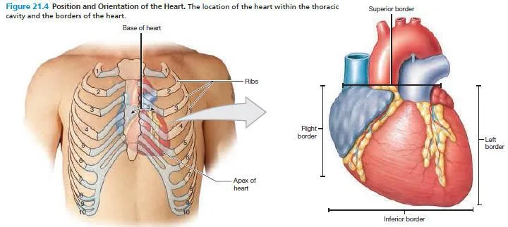
16 Orientation and Superficial Anatomy of the Heart
The heart is located within the mediastinum , between the two lungs. Because the heart lies slightly to the left of midline, the cardiac notch within the medial surface of the left lung is deeper than the cardiac notch in the medial surface of the right lung. The base of the heart is the broad, superior portion of the heart, where it is attached to the major arteries and veins of the systemic and pulmonary circuits. The base of the heart begins at the origins of the major vessels and the superior surfaces of the two atria. Thinking back to our balloon analogy, the base of the heart corresponds to your wrist (Figure 21.2b). The base sits posterior to the sternum, approximately at the third costal cartilage (Figure 21.4). The apex of the heart is the inferior, pointed tip of the heart and is formed mainly by the left ventricle. It points laterally. The apex reaches the fifth intercostal space and extends to the left of the midline.
External grooves, or sulci , of the heart show the approximate borders of the four internal chambers of the heart (Figure 21.5). A shallow interatrial groove separates the two atria. The deeper coronary sulcus marks the border between the atria and the ventricles. On the anterior surface the anterior interventricular sulcus separates the left and right ventricles. The posterior interventricular sulcus separates the left and right ventricles.
17 Internal Anatomy and Organization of the Heart
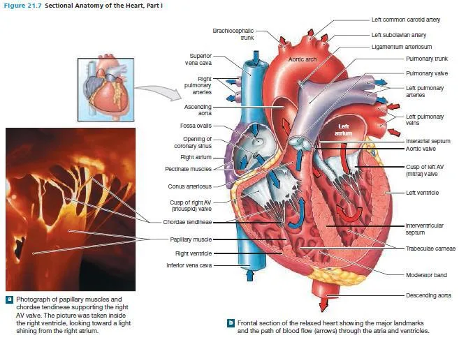
The interatrial septum (septum, wall) separates the atria, and the interventricular septum separates the ventricles. Blood flows from each atrium into the ventricle of the same side. The valves are folds of endocardium extending into the openings between the atria and ventricles. These valves open and close to prevent the backflow of blood, maintaining a one way flow of blood from the atria into the ventricles. The atria collect blood returning to the heart and then deliver it to the attached ventricle see Figure 21.7.
18 The Right Atrium
Oxygen-poor (deoxygenated) venous blood travels from the right atrium into the right ventricle. In doing so, the blood passes through an opening guarded by three fibrous flaps. These flaps, or cusps, form the right atrioventricular (AV) valve , or tricuspid valve (Figures 21.7 and 21.8). On one side, the cusps are attached to the cardiac skeleton of the heart. Their free edges are attached to connective tissue fibers called chordae tendineae . These fibers arise from the papillary muscles — cone-shaped muscular projections of the inner surface of the right ventricle. The chordae tendineae limit the movement of the cusps when the valve closes. This prevents backflow of blood from the right ventricle into the right atrium. The superior end of the right ventricle tapers to the conus arteriosus , a smooth-walled, cone-shaped pouch. The conus arteriosus ends at the pulmonary valve (pulmonary semilunar valve) . This valve consists of three thick semilunar (half moon–shaped) cusps. As blood is pumped out of the right ventricle, it passes through this valve and enters the pulmonary trunk. The pulmonary trunk is the first vessel of the pulmonary circuit. The pulmonary valve prevents the backflow of blood into the right ventricle when that chamber relaxes. From the pulmonary trunk, blood flows into both the left and right pulmonary arteries (Figure 21.5). These vessels branch repeatedly within the lungs before supplying the pulmonary capillaries, where gas exchange occurs.
19 The Left Atrium
Oxygen enters the bloodstream at the pulmonary capillaries. The oxygen-rich (oxygenated) blood flows from the pulmonary capillaries into small veins. These ultimately unite to form four pulmonary veins, usually two for each lung. These left and right pulmonary veins empty into the posterior portion of the left atrium (Figures 21.5, and 21.7b). As blood flows from the left atrium into the left ventricle, it passes through the left atrioventricular (AV) valve , also known as the bicuspid valve or mitral valve . This valve has two cusps compared to the three seen in the right AV valve. The left AV valve permits the flow of oxygen-rich blood from the left atrium into the left ventricle, but prevents blood flow in the reverse direction.
20 The Left Ventricle
The wall of the left ventricle is approximately three times thicker than the wall of the right ventricle. Contractions of the left ventricle must produce enough pressure to push the blood through the entire systemic circuit. The right ventricle, in contrast, has a relatively thin wall. It only has to develop enough pressure to push blood to the lungs and then back to the heart, a total distance of only about 30 cm (1 ft.). The internal organization of the left ventricle closely resembles that of the right ventricle (Figure 21.7). Blood leaving the left ventricle passes through the aortic valve (aortic semilunar valve) into the ascending aorta . The arrangement of the cusps in the aortic valve is similar to that in the pulmonary valve. Small, saclike dilations of the base of the ascending aorta occur next to each cusp of the aortic valve. These sacs, called aortic sinuses , prevent the individual cusps from sticking to the wall of the aorta when the valve opens. The right and left coronary arteries , which deliver blood to the myocardium, originate at the aortic sinuses. The aortic valve prevents the backflow of blood into the left ventricle once it has been pumped out of the heart and into the systemic circuit. From the ascending aorta, blood flows into the aortic arch and then into the descending aorta (Figures 21.5 and 21.7b).
21 Structure of the Heart Wall
The wall of the heart is composed of three layers (Figure 21.3):
- An outer epicardium
- A middle myocardium
- An inner endocardium
1. Epicardium: It is the visceral layer of the serous pericardium which covers the surface of the heart.
2. Myocardium: The myocardium is cardiac muscle tissue that forms the atria and ventricles.
3. Endocardium: The endocardium covers the inner surfaces of the heart , including those of the heart valves.
22 The Pericardium
The pericardium surrounds the heart and is composed of two parts:
- An outer fibrous pericardium
- An inner serous pericardium (Figure 21.2b)
The potential, fluid-filled space between these two serous layers is the pericardial cavity (Figure 21.2b–d). The pericardial cavity normally contains up to 50 mL of pericardial fluid , secreted by the pericardial membranes. This fluid acts as a lubricant, reducing friction between the opposing visceral and parietal surfaces as the heart beats.
23 Blood circulations
The circulation of the blood within the cardiovascular system can be distinguished into 3 types of circulations, which are communicating together. These are:
24 Blood vessels
The cardiovascular system is a closed system that circulates blood throughout the body. Blood vessels can be divided into Arteries & veins and capillaries . The pulmonary circuit supplies the lungs, and the systemic circuit supplies the rest of the body. The heart pumps blood into the pulmonary and systemic circuits simultaneously.
The pulmonary circuit begins at the pulmonary valve and ends at the entrance to the left atrium. Pulmonary arteries branch from the pulmonary trunk and carry blood to the lungs for gas exchange.
The systemic circuit begins at the aortic valve and ends at the entrance to the right atrium. Systemic arteries branch from the aorta and distribute blood to all other organs for nutrient, gas, and waste exchange.
After blood vessels enter an organ, further branching occurs, forming smaller and smaller blood vessels.
All chemical and gaseous exchange between the blood and interstitial fluid takes place across capillary walls. Tissue cells rely on capillary diffusion to obtain nutrients and oxygen and remove metabolic wastes. Blood leaving the capillary networks enters a network of small veins that gradually merge to form larger vessels. These larger vessels ultimately drain into either the pulmonary veins (pulmonary circuit) or the inferior or superior vena cava (systemic circuit).
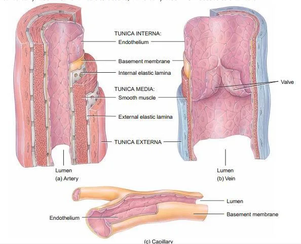
25 Structure of blood vessels
The walls of arteries and veins contain three distinct layers, from superficial to deep:
- an outer ( adventitia )
- A middle ( media )
- An inner ( intima )
The middle layer contains concentric layers of smooth muscle tissue supported by a framework of loose connective tissue. The smooth muscle cells of the media encircle the lumen of the blood vessel. When stimulated by the sympathetic branch of the autonomic nervous system, these smooth muscles contract, reducing the luminal diameter of the blood vessel. This process is called vasoconstriction . Relaxation of the smooth muscles increases the diameter of the lumen, a process called vasodilation . Any change in vessel diameter affects both blood pressure and blood flow.
26 Distinguishing arteries from veins
- Vessel walls: The walls of arteries are thicker than those of veins. The media of an artery contains more smooth muscle and elastic fibers than a vein.
- Vessel lumen: The lumen of an artery appears smaller than an accompanying vein. Arteries retain their circular shape in histological sections. In contrast, cut veins collapse, and in a histological section they often look flattened or distorted.
- Valves: Veins typically contain valves - internal structures that prevent the backflow of blood toward the capillaries. Arteries do not have valves.
27 Arteries
- They are elastic vessels, which carry blood away from the heart .
- They are branching so that; a big artery gives medium-sized arteries, which in turn give small-sized arteries, arterioles, small arterioles and finally arterial capillaries.
There are 3 kinds of arteries (according to size and function):
- Elastic arteries are the largest arteries e.g. aorta and other nearby branches. They contain a large amount of elastic tissue, which enables them to expand as blood enters their lumen from the contracting heart.
- Muscular arteries are medium-sized arteries e.g. arteries of the limbs. They contain abundant smooth muscle fibers, which allow them to regulate blood flow by vasoconstriction or vasodilatation.
- Arterioles are small arteries. Most arterioles contain considerable smooth muscles. The smallest arterioles consist of endothelium surrounded by a single layer of smooth muscle. Arterioles regulate the flow of blood into capillaries by vasoconstriction and vasodilatation.
Arteries may communicate together, forming " arterial anastomosis ", through which the blood can find an alternative channels if the main pathway is obstructed.
- Anastomosis is rich in the regions where movements can interfere with continuous constant circulation e.g. around knee and elbow.
- The arteries which have no communications with the neighboring arteries are called " end arteries " e.g. coronary arteries.
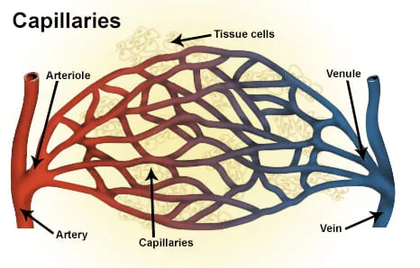
28 Capillaries
Capillaries are microscopic and most delicate blood vessels. They is the only blood vessels whose walls allow the exchange of nutrients and wastes between the blood and the surrounding interstitial fluids. Because the walls are thin, the diffusion distances are short. As a result, the exchange between the blood and interstitial fluids occurs quickly. In addition, blood flows slowly through capillaries, allowing sufficient time for diffusion or active transport of materials across the capillary walls. Some substances cross the capillary walls by diffusing across the endothelial cell lining. Other substances pass through gaps between adjacent endothelial cells. The fine structure of each capillary determines its ability to regulate the two-way exchange of substances between blood and interstitial fluid. A typical capillary wall consists of one to three endothelial cells sitting on a delicate basil lamina. The average luminal diameter of a capillary is only 8 µm, close to that of a single red blood cell.
29 Four mechanisms are responsible for the exchange of materials across the walls of capillaries and sinusoids:
- Diffusion across the capillary endothelial cells (lipid-soluble materials, gases, and water by osmosis)
- Diffusion through gaps between adjacent endothelial cells (water and small solutes; larger solutes in the case of sinusoids)
- Diffusion through the pores in fenestrated capillaries and sinusoids (water and solutes)
- Vesicular transport by endothelial cells (endocytosis at luminal side, exocytosis at basal side), water, and specific bound and unbound solutes.
30 Capillary Beds
Capillaries are the site of nutrient, waste, O2, and CO2 exchange with interstitial tissues. A collection of capillaries is called a capillary bed . A single arteriole gives rise to dozens of capillaries, which empty into several venules Fig 22.3.
31 Veins
- Wide, thin-walled vessels, which carry blood towards the heart .
- Their walls are thinner than those of corresponding arteries because the blood pressure in veins is lower than in arteries.
- Veins collect blood from all tissues and organs and return it to the heart.
- Many veins, especially those in the limbs, have valves , formed from folds of the tunica intima that prevent the backflow of blood.
- They are either superficial or deep .
- The deep veins accompany the arteries, so that each artery is accompanied by a vein but, in some parts the artery is accompanied by 2 veins " venae comitantes ".
- Like the arteries, they have tributaries.
- Venous capillaries collect into venules → small veins and finally → large veins.
- Veins are classified as large veins, medium-sized veins and venules.
32 Venous Valves
The blood pressure in venules and medium-sized veins is so low that it cannot overcome the force of gravity. For example, when you are standing, blood returning from your feet must overcome the pull of gravity to ascend to your heart. In the limbs, medium-sized veins contain one-way valves that form from infoldings of the intima (Figure 22.4).
These valves act like the valves in the heart, preventing the backflow of blood. Valves compartmentalize the blood within the veins, dividing the weight of the blood between the compartments.
As long as the valves function normally, any movement in the surrounding skeletal muscles squeezes the blood toward the heart. This mechanism is called a skeletal muscle pump .
33 Connections between arteries and veins
- Capillaries: Are microscopic blood vessels with extremely thin walls. They are lined with single layer of endothelium. Capillaries penetrate most body tissues forming network called capillary beds. The thin walls of the capillaries allow the diffusion of O2 and nutrients out of the capillaries, while allowing CO2 and wastes into the capillaries Fig 22.3.
- Sinusoids: Are similar to capillaries in that they are thin-walled blood vessels, but they have irregular and wider spaces than capillaries. They are seen in many sites e.g. liver, spleen, bone marrow and suprarenal gland. The cells lining the sinusoids include phagocytic cells Fig 22.3.
- Arterio-venous anastomosis (shunts): Are direct connections between small arterioles and small venules without the intervention of the capillaries. They are numerous in the in the skin of lips, nose, tips of the fingers and toes, intestinal mucosa and in the cavernous tissues of the sex organs. Their walls are surrounded by sphincters which open and close controlling the blood supply to the involved organs Fig 22.3.
34 Major arteries
Aorta: Arises from the left ventricle of the heart. It has three parts:
- Ascending aorta: as it ascends from the left ventricle. It gives: Right coronary artery
- Left coronary artery
- Arch of aorta: as its shape is like an arch. It gives: Brachiocephalic artery (right common carotid , subclavian arteries)
- Left common carotid artery
- Left subclavian artery
Each common carotid artery divides into:
- Internal carotid (for the brain)
- External carotid (for the face and neck)
Each subclavian artery enters the upper limb Fig.22.14.
Descending aorta: as it descends downwards, firstly in the thorax " thoracic aorta " then, it pierces the diaphragm at level of 12th thoracic vertebra and enters into the abdomen where is called " abdominal aorta ".
- ➢ Thoracic aorta: gives bronchial arteries to the lungs, branches to trachea, oesophagus and intercostal spaces.
- ➢ Abdominal aorta: gives Single branches to the GIT, Celiac trunk, Superior mesenteric artery, Inferior mesenteric artery
- Paired branches: Renal arteries to the kidneys, Gonadal (testicular or ovarian) arteries
Finally the aorta divides into two common iliac arteries (right and left) at the level of 4th lumbar vertebra. The common iliac arteries divide into two branches internal iliac and external iliac Fig.22.14.
35 Pulmonary trunk
It arises from the right ventricle of the heart.
It gives right and left pulmonary arteries which enter the right and left lungs.
36 The upper limb Arteries:
The subclavian artery continues in the upper limb and as it enters the axilla, it is called axillary artery . The later continues in the upper arm as brachial artery . The brachial artery divides into radial and ulnar arteries in front of the elbow joint.
37 The lower limb Arteries:
The femoral artery gives branches in the thigh and then goes behind the knee joint and here it is called popliteal artery . Popliteal artery divides into anterior tibial and posterior tibial arteries in the leg.
38 Major veins
Superior vena cava (SVC): It drains the venous blood from head and neck, upper limbs and thorax . It is formed by the union of right and left innominate veins. Each innominate vein is formed by of subclavian vein (continuation of the axillary vein) and internal jugular vein (from the head and neck).
Inferior vena cava (IVC): It drains the venous blood from all the body below the diaphragm . It is formed in the abdomen by the union of right and left common iliac veins at the level of 5th lumbar vertebra. It ascends on the right side of the aorta, pierces the diaphragm and enters the thorax and finally opens into the right atrium of the heart Fig.22.20.
39 Superficial veins of upper limb:
Superficial veins of upper limb are used for venipuncture and blood transfusion . The constant position of the cephalic vein makes it available even if not seen as in obese patients.
- 1- Basilic vein begins from the medial side of the hand and ascends up in the forearm and finally in the upper arm where it becomes as the axillary vein.
- 2- Cephalic vein begins from the lateral side of the hand and ascends up in the forearm and finally in the upper arm.
- 3-The median cubital vein is a communication between the basilica and cephalic veins in front of the elbow joint. This vein is used for giving intravenous injections Fig 22.20.
40 Nursing Uganda Clinical Lens
Use Cardiovascular system as a practical nursing topic, not only a memorized definition. Prioritize airway, breathing, circulation, pain, asepsis, wound healing and early complication detection.
- What to understand first: define cardiovascular system, identify the normal or expected pattern, then explain what changes when the patient is unwell.
- Why it matters in care: the nurse must recognize risk early, explain findings clearly, document accurately and know when to escalate.
- How to revise it: connect each point to assessment, nursing diagnosis or care problem, intervention, rationale and evaluation.
41 Assessment Guide
- Vital signs, pain, bleeding, perfusion, level of consciousness and injury pattern.
- Wound appearance, drainage, odour, swelling, temperature and surrounding skin.
- Fluid balance, mobility, nutrition, surgical site risk and ordered investigations.
42 Nursing Priorities, Rationales and Outcomes
- Stabilize urgent problems first, then prepare for investigations or theatre care.
- Maintain aseptic technique, pain control, wound care and documentation.
- Prevent shock, infection, pressure injury, deep vein thrombosis and delayed healing.
The rationale for these priorities is patient safety: nursing actions should prevent deterioration, reduce discomfort, support recovery and create clear evidence for the next caregiver.
- Expected outcome: The patient remains stable, wound healing progresses, pain is controlled and complications are recognized early.
43 Patient Teaching and Revision Check
- Explain cardiovascular system in simple language the patient or caregiver can repeat back.
- Teach warning signs, medicine or follow-up instructions, hygiene or lifestyle points where relevant.
- For exams, prepare a short answer using: definition, causes or risk factors, signs, assessment, management, complications and prevention.
- For ward practice, document baseline findings, actions taken, patient response and the plan for review.
Illustrations and Diagrams (21)

13 more diagrams available — open the lesson for full illustrations.
Related Video Lectures
Watch nursing lecture videos on YouTube for this topic. Opens in a new tab.
Watch on YouTubeExternal link: YouTube may use its own cookies and terms. Nursing Uganda is not affiliated with YouTube.